Tools & Approaches
The Wanner team combines imaging and biochemical approaches at the intersection of structural and molecular neuroscience with direct translational use in the clinic.
One rat, one number:
Histopathology pipeline algorithm to quantify brain injury.
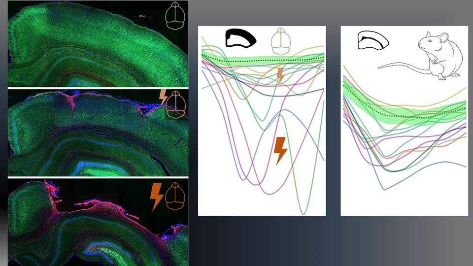
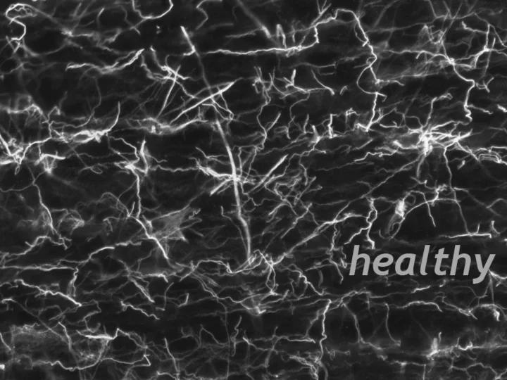
Neuronal landscape from healthy to injured rat brains: Top micrograph show healthy rat cortex with bright neuronal stain (Neurofilament light, NFL, green). Middle/bottom depict 1 month postinjury cortical contusion with neurodegeneration (NFL loss) and glial scarring (GFAP, red). Mediolateral traces of NFL loss for each injured rat contrast with normal averages (dotted green line with green variance band). Comparisons span both cortical gray matter (center) and corpus callosum white matter (right).
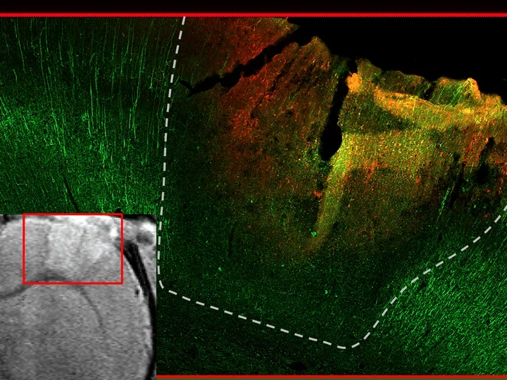
From voxels to tissue:
MRI meets microscope in validating rat brain trauma.
Internal validity is documented by aligning in vivo structural MRI hyperintensity (courtesy Neil Harris, UCLA) with postmortem acute loss of neuronal fiber signal (Neurofilament light, green) around bleeds in the contused core visualized on immunofluorescence stained histological sections that are co-registered with one another in the same rat six hours after injury.
Microscopy and Imaging
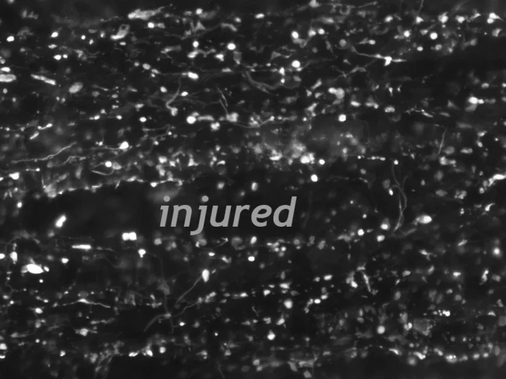
The Wanner lab established creative microscopic imaging tools and quantitative analyses using confocal microscopy, time-lapse imaging and ultrastructural studies. We measure biomarker changes in single cells, quantify GFAP filament aggregation, local protein translation with subcellular resolution and determine the extent of cellular injury including astrocyte fiber beading, called clasmatodendrosis, and cell body swelling, called cytotoxic edema after trauma.
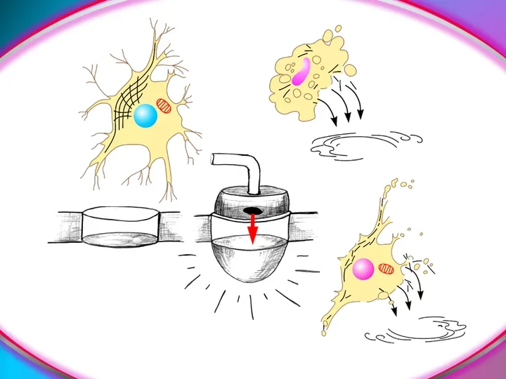
Culture models:
Human culture model reveals how brain cells respond to whiplash trauma
Human neocortical cells on flexible membrane endure sudden impacts that resemble traumatic injury. Sheared, wounded cells are monitored through automated microscopy and released biomarkers are assayed in surrounding fluids, offering unique insights into cytological and matching biomarker trauma dynamics.