Microscopy and Imaging


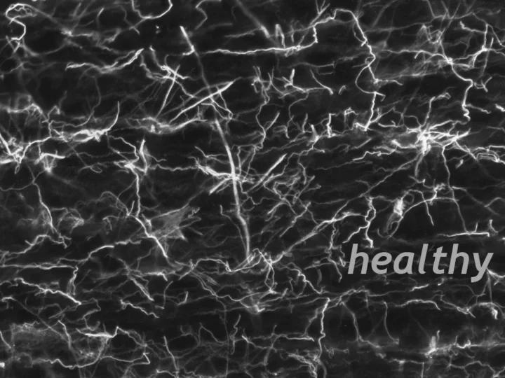
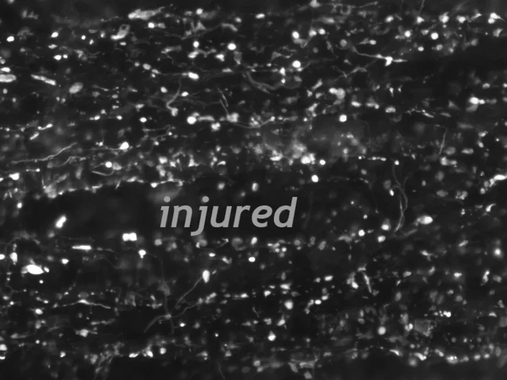
The Wanner lab established creative microscopic imaging tools and quantitative analyses using confocal microscopy, time-lapse imaging and ultrastructural studies. We measure biomarker changes in single cells, quantify GFAP filament aggregation, local protein translation with subcellular resolution and determine the extent of cellular injury including astrocyte fiber beading, called clasmatodendrosis, and cell body swelling, called cytotoxic edema after trauma.